
If it feels like dental visits involve more photography than they used to, you're not imagining it. Between X-rays, the little wand camera that takes close-ups inside your mouth, and photos of how your teeth come together, a modern checkup can generate quite a few images.
It's fair to wonder why. Here's what all those pictures are actually for — and why they're one of the more useful things a dental practice can do for the long-term health of your teeth.
X-rays Are Only Part of the Picture
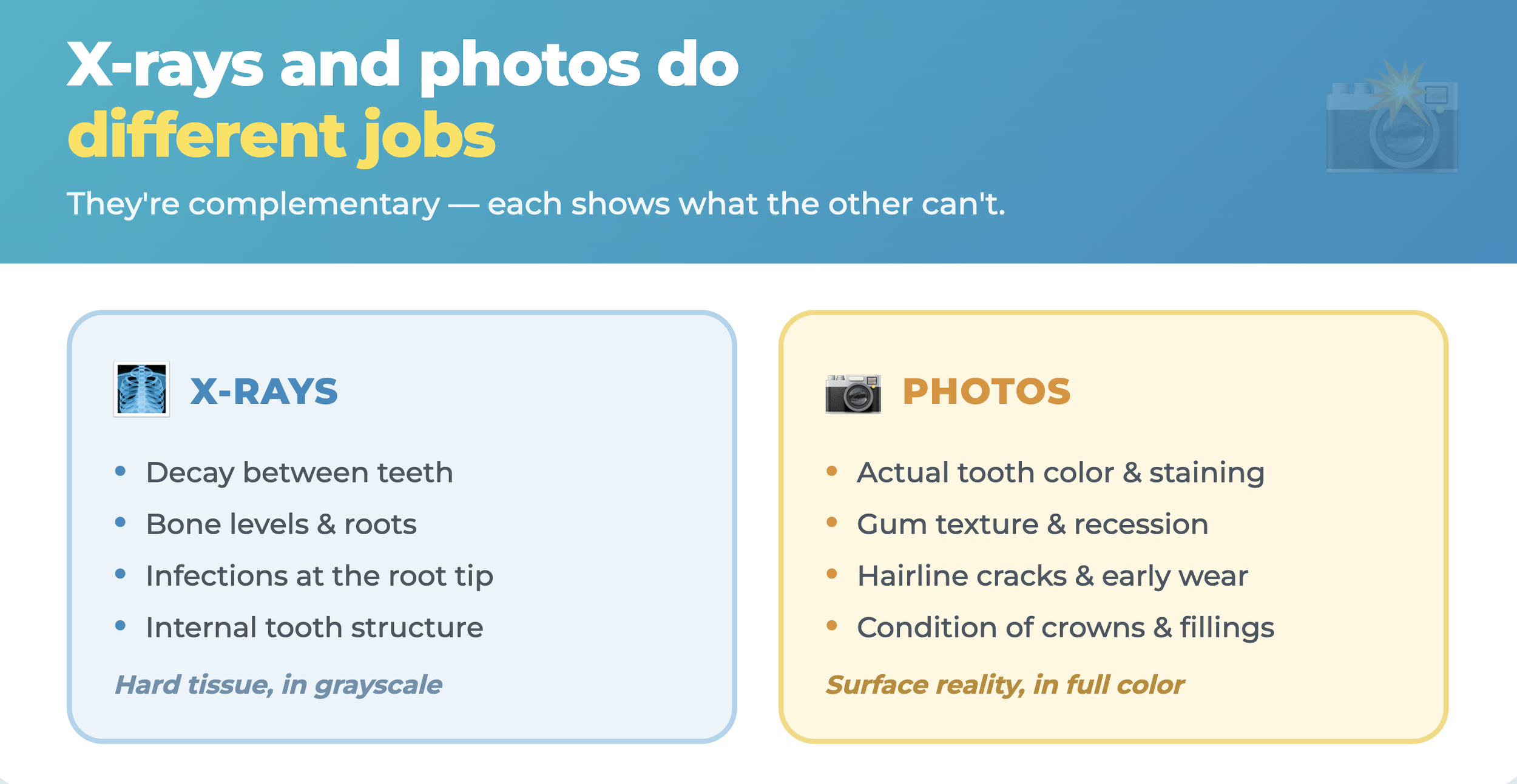
When patients think about dental imaging, X-rays are usually what comes to mind. And X-rays are essential — they show what the eye can't see: decay between teeth, bone levels, the roots, infections at the root tip, and the internal structure of each tooth.
But X-rays have a specific limitation: they show hard tissue in shades of gray, not the surface reality of your mouth in full color and detail. They can't show you the actual color of a tooth, the texture of your gums, the early wear pattern on a chewing surface, or the hairline crack running across the enamel. That's where the other kinds of dental photography come in — and why the two work together rather than one replacing the other.
What Intraoral and Bite Photos Actually Capture
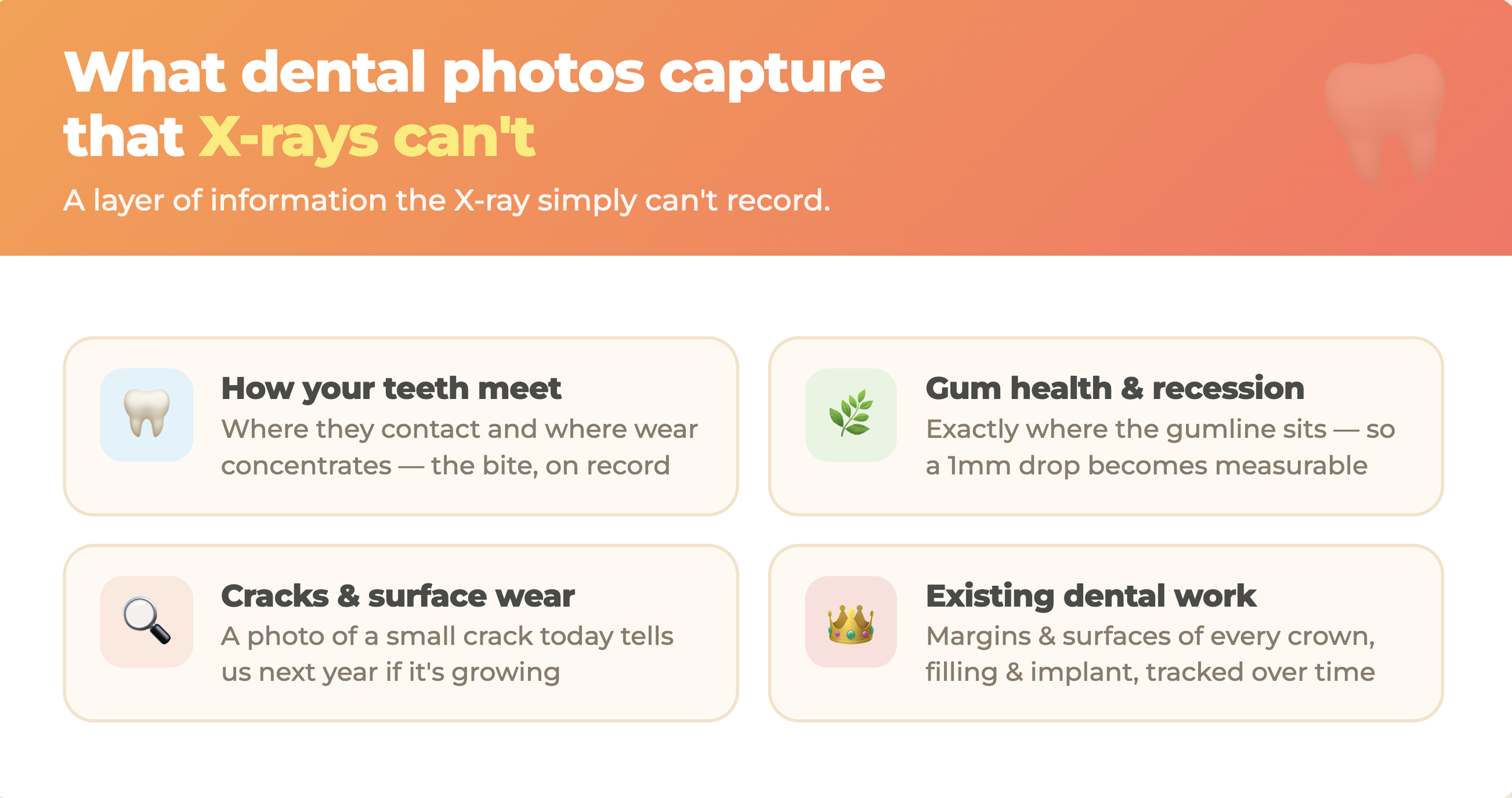
The close-up photos taken with a small intraoral camera, along with photos of how your teeth meet, record a layer of information X-rays simply can't.
How your teeth come together (occlusion).
Photos of your bite document how the upper and lower teeth meet — which surfaces contact, where wear is concentrated, and how the relationship sits at this moment in time. Since the bite changes gradually as teeth wear, shift, and get restored, having it on record matters.
Gum health and recession.
The color, texture, and position of your gum tissue tell a detailed story about periodontal health — and much of it is visual, not radiographic. Photographs capture exactly where the gumline sits on each tooth, so that recession of even a millimeter or two becomes measurable against a previous image.
Cracks, wear, and surface changes.
Hairline cracks, early enamel wear, staining changes, and the condition of existing fillings and crowns are all surface findings that live in the visual record, not the X-ray. A close-up photo of a small crack today is the reference point that tells us, a year from now, whether it's growing.
The condition of existing dental work.
Photos document the current state of every crown, filling, and implant — their margins, their surfaces, any early signs of wear or failure — so changes can be tracked precisely over time.
The Real Value: Comparison Over Time
Here's the single most important reason for all the photography, and it's worth understanding clearly: most dental problems develop slowly, and the most powerful diagnostic tool is comparison against the past.
A tooth doesn't usually go from healthy to needing a root canal overnight. Gums don't recede all at once. Cracks don't appear fully formed. Wear doesn't happen in a day. These changes unfold gradually — often so gradually that at any single appointment, they look like "normal." The change only becomes visible when this year's image sits next to last year's.
That's what a good photographic record makes possible. A gumline that dropped a millimeter. A crack that lengthened slightly. A filling whose margin started to break down. A spot of wear that's deepening. Individually, at one visit, each might be easy to overlook. Side by side across time, the trend is obvious — and catching a trend early is what allows a small, simple intervention instead of a large, complicated one later.
Without the record, we're relying on memory and a single moment. With it, we're tracking your mouth like a series of data points, watching for the direction of change rather than just its current state.
It Helps You See What We See
There's a second real benefit, and it's about you, not just the clinical record.
Dental findings can be hard to picture when they're only described in words. "There's some recession on the lower left" or "this crown's margin is starting to break down" means far more when you're looking at a clear, magnified photo of exactly that. Being able to see your own mouth the way we see it — the crack, the wear, the inflammation, the change from last year — transforms the conversation from something you're asked to take on faith into something you can evaluate yourself.
That matters for real decisions. When it's time to weigh whether to treat something now or watch it, seeing the actual image — and ideally the comparison to how it looked before — helps you make a genuinely informed choice about your own care. Patients who can see what's happening tend to understand their options better and feel more confident in what they decide.
Frequently Asked Questions
Why does the dentist take so many pictures?
Because most dental problems develop slowly, and comparing images over time is the most reliable way to catch small changes early. Photos capture surface details X-rays can't — gum position, cracks, wear, and the condition of existing dental work — and create a record to track against future visits.
What's the difference between dental X-rays and photos?
X-rays show hard tissue and internal structures in grayscale — decay between teeth, bone levels, roots, and infections. Photos capture the color, texture, and surface reality of teeth and gums. They're complementary: each shows what the other can't.
What is an intraoral camera?
A small wand-shaped camera that takes magnified close-up photos inside your mouth. It documents cracks, wear, gum condition, and existing dental work in detail, and lets you see those findings on a screen alongside the dentist.
Do dental photos expose me to radiation?
No. Intraoral and bite photographs use ordinary light, not radiation — they're simply photographs. Only X-rays involve radiation, and modern digital X-rays use very low doses.
How do photos help catch problems early?
By making gradual change visible. A crack, a receding gumline, or a breaking-down filling margin may look normal at any single visit. Compared against an image from a year ago, the change becomes obvious — early enough for a simpler fix.
The Thing About Slow Change
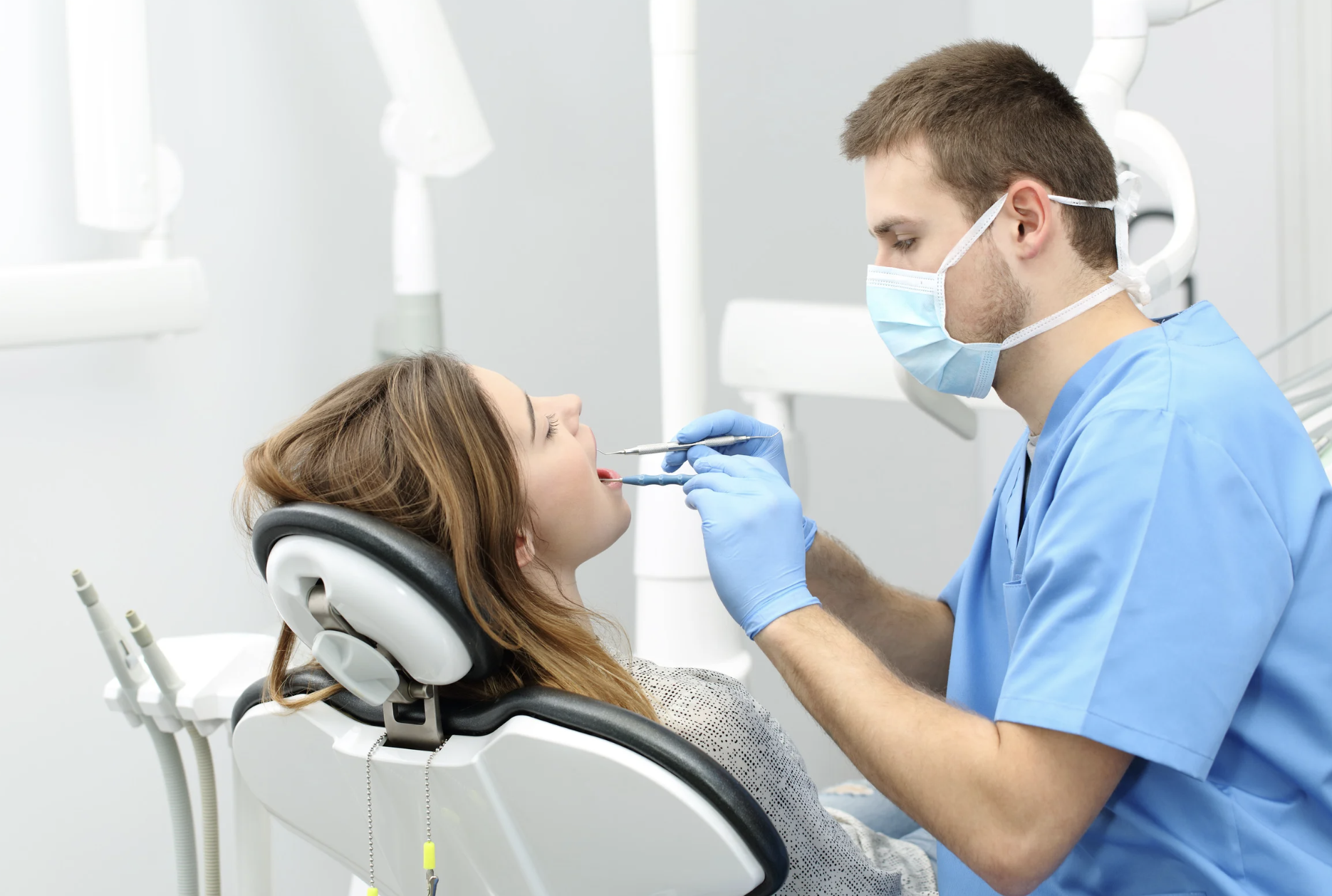
Almost everything that goes wrong with teeth happens gradually. That's easy to forget, because we tend to notice dental problems only at the moment they finally start to hurt or show — which is usually late in a process that began quietly much earlier.
Photography is how a dental practice fights that. It turns a series of separate appointments into a continuous record, so that slow change has somewhere to show up before it becomes a problem you can feel. That's not extra fuss — it's the difference between finding things early and finding them late.
So the next time there's a camera in the mix at your visit, know that it's doing real work: building the record that helps keep small things small. If you're overdue for a checkup and haven't had a good set of images taken in a while, give us a call — a current baseline is one of the most useful things your teeth can have.
📍 State Avenue Dental Office — Kansas City, KS (KCK) 🗣 English • Korean • Spanish